Introduction

Wilhelm Röntgen discovered X-ray November 8th 1895, when he did experiments with cathode rays in a vacuum tube. He worked in a dark room and the tube was covered with black opaque paper. However, on the standing nearby screen, covered with fluorescent barium tetracyanoplatinate, Röntgen saw a glowing spot. Wilhelm placed his hand between the tube and the screen, on the path of the invisible light, and on the screen he saw a strange shadow of his hand: bones were clearly visible and there were only vague shapes of soft tissues.
To capture and save images of the shadows from the X-rays, he used ordinary photoplates. Fortunately, sensitive to visible light silver based photoemulsions turned out to be sensitive to the X-ray too. These photoplates became the first X-ray detectors, and Wilhelm made the first radiograms more than 100 years ago with them.
More than 100 years of scientific progress led to the creation of a number of various detectors for recording X-ray images. Developments of the microelectronics and semiconductor manufacturing technologies are crucial for development of the modern X-ray detectors. These detectors can transform the energy of the X-ray photon directly to the electrical signal. They allow capturing detailed, digital, high-resolution X-ray images.
Digital images are easy to work with. For example one can merge multiple macro images into an image of the whole object and represent monochrome images in false colors like Simon Procz did with this X-ray image of a flower he did in 2012.

Simon Procz captures these X-ray images using hybrid pixel detectors, specifically the Medipix detectors. The technology of the hybrid pixel detectors was developed in CERN for use in the main detector experiments of the Large Hadron Collider. Thanks to the Knowledge Transfer program this technology was shared to be used in practical and commercial use.
Hybrid Pixel Detectors
The “hybrid“ these detectors got because they are composed of two main parts: semiconductor sensor and readout ASIC. The two parts of the detector are separate and can be developed and optimized independently.
Hybrid pixel detectors owe their existence to the developments of microelectronics and semiconductor technologies. The sensor and the readout ASIC are connected by bump bonding and flip-chip technology, which was developed in the 60s for assembling ICs at IBM.
The sensor of a hybrid pixel detector can be made from various semiconductor materials, depending on the required absorption and electrical properties. The silicon sensors are manufactured from the monocrystalline silicon wafer using technologies of the semiconductor industry.
The detectors are “pixel” because the sensor is split into the square pixel cells and each pixel corresponds to the signal measurement unit in the readout ASIC.
Hybrid pixel detectors convert the energy of a photon into free charges in the semiconductor which directly forms the useful electrical signal. This type of detectors are also called Photon Counting detectors, as they allow detecting a single photon. Older types of X-ray detectors, scintillation detectors, transform the photon energy in two steps. They use scintillating crystals to first transform an X-ray photon into a number of visible spectrum photons. These visible photons are then detected with a regular CCD or CMOS photosensor array, which is also split into pixel elements. Scintillation detectors work with a semiconductor photosensor array in the integrating mode, collecting the energy over some time, as they are not sensitive enough. Hybrid pixel detectors have better sensitivity, energy and space resolution thanks to the direct energy conversion.

Medipix project
The Medipix project was born in CERN as one of the Knowledge Transfer programs. Its goal is to share the technologies developed first for scientific research for commercial and practical use.
The first generation of the detectors - Medipix1 - was presented in 1997. The pixel size is 170 by 170 um and the sensitive matrix is 64 by 64 pixels. The readout ASICs were manufactured using the 1000 nm Self Aligning CMOS technology. The detector is able to measure the signal as low as 2000 electron charges. The signal from the pixel was digitized by the comparator with a configurable threshold with 3-bit resolution. The events are counted by the 15-bit counter. The data from the detector is read with a special 16-bit bus with Muros1 protocol. The clock frequency for data transfer was 10 MHz so one frame is read out in 384 us.
The project for developing the next generation - Medipix2 - started in 1999. The matrix is 256 by 256 pixels and each pixel is 55 by 55 um in size. Readout ASICs are manufactured with the 250 nm CMOS technology. The novelty in the Medipix2 detectors is in implementing an energy window for signals with two comparators for lower and higher bounds for energy. Within the Medipix2 project there are variants of detectors. The Timepix detectors first presented in 2005 measure not the number of events but time the signal is above the threshold. This time corresponds to the particle energy. For dosimetry the Dosepix detectors were developed. And VELOpix detectors are used in the LHCb experiment.
The third generation project - Medipix3 - started in 2005. The matrix and pixel sizes are configurable. Matrix can be 128 by 128 pixels with pixel size 110 by 110 um or 256 by 256 pixels with pixel size 55 by 55 um. The readout ASICs are manufactured with 130 nm CMOS technology. The main feature of the Medipix3 is solving the problem with the noise due to charge sharing between pixels.
The Medipix4 project started in 2016. The goal of the project is to develop 4-way tileable detectors by using Through Silicon Via technology. Previous detectors could be tiled only from 3 sides due to wirebond connection to the PCB.

Working principle
The working principle of semiconductor based detectors is based on the generation of free electron-hole pairs in the semiconductor when the energy of a photon or another particle is absorbed. Due to the reverse bias voltage applied to the sensor, newly created electron-hole pairs are moving and electric current is flowing through the sensor. This current is measured by the readout ASIC. The number of created free charges is proportional to the energy of a photon, and so does the magnitude of the electric current.

Semiconductor sensor
The sensor is made of semiconductor material and transforms the particle energy into the electrical signal. The main part of the sensor is a piece of n-type semiconductor. The bottom of the sensor is covered by some insulating material and metallic bump-bond pads in a 2D grid. Above each pad a small amount of p-type semiconductor is implanted in the sensor. Each element of a sensor matrix is thus a diode. The top of the sensor is covered by a thin aluminium layer, which is used to apply reverse bias voltage.

The reverse bias voltage increases the depletion region of the p-n junction to almost all sensor volume. In the depletion region the electrons and holes are recombined and the concentration of free charges is very low.

Interactions of X-rays with sensor
X-rays are electromagnetic radiation with wavelengths from around 10^2 to 10^-3 nm, which corresponds to the energies of a photon from 10 eV to 1 MeV. In medical and industrial applications diagnostic devices use relatively soft X-ray sources with the wavelengths of radiation in the range from around 10 to 200 keV.
The X-ray photon can be detected only if it transfers its energy into the sensor material partially or in full. For soft X-rays the main energy transfer mechanisms are photoelectric effect and Compton scattering. If the photon energy is higher than 1.022 MeV the electron-positron pair production mechanism starts occurring.
Photoelectric effect results in full energy transfer from the photon to ionization of the atom and kinetic energy of the free electron. There are secondary interactions - fluorescence of a new secondary photon and Auger effect which creates an additional free electron.
In the case of Compton scattering only part of the photon’s energy is used for ionization and afterwards the photon changes its direction.
There is also a mechanism which only changes the direction of the photon without it losing energy - Rayleigh scattering.
Secondary photons and scattered photons can again interact with the atoms of the sensor. The interaction cascade will create new photons and free charges until all the energy of the initial X-ray photon is used up. However the spatial information becomes blurred and some of the energy can be lost when secondary photons leave the sensor pixel to other pixels or entirely from the detector.

The absorbed energy should be roughly 3 times higher than a band gap to create a free electron-hole pair in a semiconductor. Number of free charges created is proportional to absorbed energy. Silicon has a bandgap of 1.12 eV. An X-ray photon with energy around 30 eV in case of total absorption will generate in a silicon sensor around 10000 free charges.

Selecting the material for the sensor is a typical engineering tradeoff problem. For spectroscopic detectors the preferred materials are semiconductors with higher probability of photoelectric effect. This means the semiconductor should have a higher atomic number. But with a higher atomic number higher the rate of secondary photon generation, so the spatial information and the photon’s trajectory information is blurred. Particle trajectory tracking detectors can use lower atomic number materials and spare energy loss out of pixel to keep the spatial information. Semiconductors with a lower band gap, e.g. germanium with 0.5 eV, are more sensitive as more free charges are generated from the photon’s energy. But they will have more thermal noise and must be actively cooled to work effectively. In the medical field X-rays are relatively soft and higher atomic number sensors have to be used. The examples of sensor materials for medical X-ray are cadmium telluride CdTe or gallium arsenide GaAs.
Here the hybrid architecture of the Medipix and similar detectors comes into play. The same readout ASIC can be used with different sensors for different tasks. Sensors can also have some complex internal structure. As an example this patent for a semiconductor sensor to be used with Medipix detectors from the JINR, one of the Medipix4 project members. Its feature is splitting and separating each pixel with the layer of insulator to decrease the charge blurring and sharing between pixels.
Signal formation
Due to the reverse bias voltage the newly created free electrons move towards the top aluminum layer with positive potential and holes toward the readout ASIC towards zero potential. The electric current flows through the sensor and readout ASIC measures it.
Mathematically the current signal on the electrodes from the movement of free charges in the sensor is described by the Shockley–Ramo theorem. The signal depends on the sensor geometry, bound charges distribution and mobility of free charges. The total induced charge on the electrode is proportional to the number of free charges generated inside the sensor from the photon's energy.
Readout ASIC
Readout ASICs of the hybrid pixel detectors have analog amplifiers and digital circuits corresponding to each pixel of the sensor. The signal from the sensors gets processed, digitized and recorded with these circuits.
The general schematic diagrams of the analog input amplifiers and digital circuits are shown on figures below.

The current sensitive amplifier measures the current from the sensor. Its output voltage is proportional to the integral of the sensor current due to the capacitor in the feedback loop. The integral of the current equals the total number of free charges generated in the sensor from the photon's energy. The current signals are short and can be modeled by a delta-function. The output voltage of the current sensitive amplifier is a step function. The input is a gate of a MOS transistor and input currents are low, lowering error in sensor current measurements.
To improve the signal’s time duration, frequency characteristics and SNR signal shapers are used. Usually it is a pair of a differentiating high-pass filter and an integrating low-pass filter. The simplest configuration is a pair of a CR and a RC circuits with opamps. Resulting filter is a band-pass semi-gaussian signal shaper.

After analog processing the signal is digitized. For digitization the regular analog-to-digital converters can be used, which will record the shape of the signal. In the Medipix1 detectors the digitization is done simpler - with a latching comparator with a configurable threshold. The threshold is set by the 3-bit digital-to-analog converter. This is enough to detect the X-ray photon event and assess its energy.
A 15-bit counter is incremented each time the signal is above the threshold. Closing the shutter switches the ASIC from the detection mode into the data transfer mode. In the data transfer mode the values of the counters of each pixel are transferred via the external interface.
Applications
One of the main applications for the hybrid pixel detectors is non-destructive analysis of inner structure of the objects in medicine and in industry. Interestingly it rhymes with the first application of the X-rays by Röntgen for radiography of hands and household items.
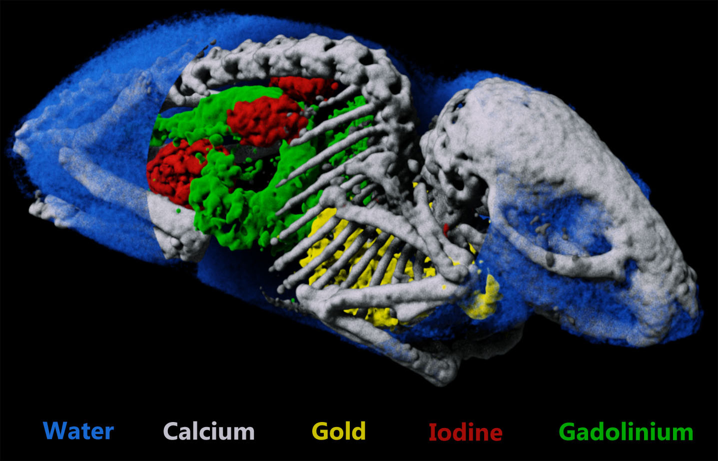
The most advanced X-ray imaging method is spectral computed tomography. It can be medical human scanners and micro-CT scanners for industrial applications. The energy measurement capabilities of the hybrid pixel detectors allow these scanners to measure the absorption spectrum of the object and determine their atomic content.

In medical CT determining the atomic content allows better differentiating between tissues, organs and contrast agents. Higher sensitivity of the photon counting detectors allow using lower intensity X-ray sources to get the images of the same quality, lowering dose received by patients.

Beside the Medipix family of detectors other companies also produce their own readout ASICs for hybrid pixel detectors. Main medical devices manufacturers develop their own technologies for future spectral CT scanners with hybrid pixel detectors to allow photon counting and spectral measurements. Philips developed ChromeAIX ASIСs and is a member of the European Spectral Photon Counting CT research project. Siemens develops readout ASICs with CEA and MC1 companies and their spectral CT scanner with photon counting capabilities has already received FDA approval. General Electric acquired Prismatic Sensors start-up for their know-hows in X-ray detector technologies.
Another application of the Medipix detectors is dosimetry. The dosimeter with Timepix detector in the form of the USB Flash drive is flying in the ISS and can be seen on some photos with astronauts. Inside the Orion spacecraft the Battery-operated Independent Radiation Detector (BIRD) with Timepix detector is installed. The Linear Energy Transfer Spectrometer (LETS) dosimeter was installed on the Peregrine lunar mission which was launched on January, 8th this year but wasn’t able to reach the Moon and burned down in the Earth’s atmosphere 10 days later. The LETS dosimeter has Timepix detectors as a main measurement unit.
Conclusions
This article is a very brief and surface level introduction to the hybrid pixel detectors technology. I’ve used a few sources, scientific papers, textbooks. It was very helpful to have a lot of CERN’s lectures open and available to everyone https://cds.cern.ch/. The list below contains the main sources I used with information about history, physics, electronics and application of X-ray detectors.
W. C. Röntgen, “On a New Kind of Rays,” Nature, vol. 53, no. 1369, pp. 274–276, 1896.
E. H. M. Heijne, “History and future of radiation imaging with single quantum processing pixel detectors,” Radiation Measurements, vol. 140, p. 106436, 2021.
D. S. McGregor and J. K. Shultis, Radiation detection: concepts, methods, and devices, 1st ed. Boca Raton: Taylor and Francis, 2020.
R. Ballabriga, “The Design and Implementation in 0.13 um CMOS of an Algorithm Permitting Spectroscopic Imaging with High Spatial Resolution for Hybrid Pixel Detectors,” Universitat Ramon Llull, 2009.
R. Ballabriga, M. Campbell, and X. Llopart, “Asic developments for radiation imaging applications: The medipix and timepix family,” Nuclear Instruments and Methods in Physics Research Section A: Accelerators, Spectrometers, Detectors and Associated Equipment, vol. 878, pp. 10–23, 2018.
R. Ballabriga et al., “Review of hybrid pixel detector readout ASICs for spectroscopic X-ray imaging,” J. Inst., vol. 11, no. 01, pp. P01007–P01007, Jan. 2016.
H. Hayashi, N. Kimoto, T. Asahara, T. Asakawa, C. Lee, and A. Katsumata, Photon Counting Detectors for X-Ray Imaging Physics and Applications. Cham: Springer International Publishing AG, 2021.